There are many case of the food poisoning which develops with hours after infection. These illnesses are caused by salmonella organisms other than S.tyhphi or S.paratyphi, for example Salmonella enteritidis. They are referred to as salmonellosis. The economic and social costs of food poisoning can be very high. Salmonellosis in the England and Wales was estimated to cost between ₤ 231 and ₤ 331 million in 1988 and 1989 in terms of lost production through sickness related absence from work, investigation and treatment.
In contrast to typhoid, salmonellosis organisms have low infectivity, in other word large numbers of bacteria are required to cause infection. This is also in contrast to the high infectivity of E. coli 157, an increasingly important agent in the food poisoning. Other bacteria may also cause food poisoning, for example Listeria Monocytogenes which is particularly associated with pate and dairy product such as soft cheese and listeriosis. Staphylococcus as soft cheese listeriosis. Staphylococcys aureus, Bacillus and Clostridium species are also well known causative agents which are beyond the scope of this contents of this blog.
Salmonella enteritidis is particularly associated with the eggs and meat from poultry. It was important in the rising incidence of salmonellosis from 1987 and accounted for 53% of salmonellosis in 1989 and 63% in 1991. At that time hen’s eggs represented a newly recognized sources salmonellosis.
How this Salmonella is transmited and what are it’s signs and symptoms
Salmonellosis is a classic food borne disease which can be easily infects the human as well as the many food animals like the poultry, pigs and cattle from which the human is directly infected. The faeces of rats, mice and domestic pets may also contaminate food human food. The sign and symptoms of the salmonella food poisoning appear suddenly, within 12 to 24 hours after eating contaminated food. Vomiting and diarrhea occur, accompanied by a fever with a high temperature. Abdominal pain or discomfort, and headache, usually occur. Most people recover within a few days.
Dehydration is a risk and may lead to complications such as low blood pressure and kidney failure. These complications are responsible for most of the death that occur. The elderly and the young, particularly babies are most at risk. Accurate diagnosis requires a culture of the faeces to isolate the organisms.
How salmonella and food poisoning can be treated and preventedThe treatment is to replace fluid and salt loss and to use drugs. Antibiotics are effective against salmonella organisms and include tetracycline and ampicillin but they do not shorten the occurs of acute diarrhea. Recently drug resistant strains of Salmonella have begun to appears. There are several methods of prevention.
Carriers: Symptom less carriers of the disease can retain the organisms in the faeces for some time and present a problem in society. About 2-5 people per thousand of the general population are thought to be carriers. Known carriers and people who have suffered the disease are not allowed to work in the food industry until samples of their faeces have been shown to be clear of the pathogenic organisms.
Hygiene in the home and food trade: Storage preparation and cooking of food should all be carried out hygienically. Food should be stored cool or out hygienically. Food should be stored cool or refrigerated to minimize bacterial growth. Great care should be taken in the kitchens to separate the different preparatory processes, so that raw meat, particularly poultry and eggs do not cross-contaminate other food stuffs. Cutting board, dish cloths, dirty kitchen utensils and unwashed hands can all harbor salmonella and bring about transfer from food to food and place to place. Environmental Health Officers regularly inspect restaurants, shops and factories.
Through cooking: Cooking should be thorough enough to kill all bacteria. One of the great dangers in not thawing frozen food sufficiently. In addition, the Chief Medical Offices recommended in 1988 that raw eggs should be avoided and vulnerable groups such as the elderly, sick, babies and pregnant women should consume only eggs which have been cooked until the white and yolk are solid.
 Meat inspection:
 by the Environment Health Officers is essential and any animals suffering from salmonellosis should not be used in the food industry. Low levels of salmonella contamination are regarded as inevitable amongst poultry and only large outbreaks result in slaughter of the birds.
Food poisoning is a notifiable disease. However many mild cases go unreported. In 1995 food poisoning from all causes was at its highest level since records began in 1949: 80000 cases were recorded in 1995 compared with 63000 for 1992. Accurate monitoring of the incidence of food poisoning is essential in helping to decide what preventative measures to take.
Control of rodents whose faeces may contain salmonella
Proper sewage disposal
Government intervention: In 1989 the government took steps to control the rise in salmonella case in Britain in salmonella cases. Testing of poultry flocks became compulsory. More than three million infected laying hens were slaughtered and the Food Safety Act of 1990 raised standards in food production.
 Annelids
  Anthropods
 Vertebrates
 The simpler animals such as cnidarians and Platyhelminths lack specialized systems for the transport and distribution of materials. The organisms in these groups possess a large surface area to volume ration and diffusion of gases over the whole body is surface is sufficient for theirs needs. Internally the distance that materials have to travel is again small enough for them to move by diffusion or Cytoplasmic streaming.

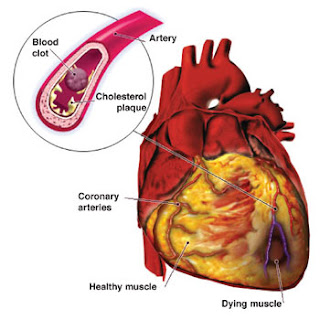
 A number of factors are known or believed to be involved in development of atherosclerosis, and hence cardiovascular disease. Some are more important than others and usually several act together to bring it about. The three most important are diet, hypertension (high blood pressure) and smoking.
 Diet: Atheroma contains fats and cholesterol, and it has been shown clearly that experimental animals fed on a high fat diet develop diseased arteries. In countries such as Greece and Japan where the average diet is relatively low in fat, cardiovascular disease is much less common. The main problem is caused by saturated fats which cause a rise in blood cholesterol levels. In 1992 the UK government recommended that the population from saturated fatty acids should be reduced from 17% (proportion in 1990) to no more than 11% by 2005 (a 35% reduction). A 12% reduction in total fat was also proposed (from about 40% to 35% of food energy). On the other hand, polyunsaturated f atty acids, found in unsaturated fats, are thought to help reduce cholesterol levels in blood and are therefore beneficial to health.
 Hypertension (High Blood Pressure): Raised blood pressure can considerably increase the chance of developing cardiovascular disease. It has been shown that men under the age of 50 years with a blood pressure of 170/100 are twice as likely to die of coronary heart disease as men with normal blood pressure of about 120/80. High blood pressure itself is associated with a number of different factors, including stress, obesity, smoking, drinking excessive amounts of alcohol and lack of exercise. There is also a genetic predisposition in some people. Some of these factors can obviously be avoided by changes in lifestyle.
 Smoking: Heavy smokers are more likely to develop cardiovascular diseases. For example, people under the age of 45 years who smoke more than 25 cigarettes a day are 15 times more likely to die of heart disease than non-smokers. Nearly 40% of cardiovascular deaths re due to smoking. Smoking increase atherosclerosis and decreases the ability to remove blood clots that build up at atheromatous plaques.
 Physical exercise: Many studies have shown that the more a person is physically active at work or during their leisure time, the less chance they have of suffering from cardiovascular disease. The effects of suffering from cardiovascular diseases.
 In the developed countries infectious diseases are no longer the major causes of death. The average life expectancy is now about 76 years. As people age however they become prone to disease of the heart and blood vessels, and cancers, and these two types of disease account for about two thirds of all deaths in developed countries.
 Myocardial refers to heart muscles; infraction means suffocation due to lack of oxygen.) If thrombosis occurs in the brain (cerebral thrombosis) a stroke may occurs. Strokes are sometimes referred to as cerebra vascular accidents. They are also caused by cerebral hemorrhage. They usually result in permanent damage to the cerebral hemispheres due to oxygen starvation. The cerebral hemispheres are the conscious part of the brain and control may functions such as speech and motor coordination. Both heart attacks and strokes may result in death.
 Thus coronary heart disease has two main forms, angina and mycocardial infraction (heart attack). A heart attack may be caused by a coronary thrombosis or simply by narrowing of the artery by atherosclerosis until the blood supply by narrowing of the artery by atherosclerosis until the blood supply is sufficiently restricted. About half a million people a year in Britain have heart attacks and about one third die as a result. Half of these die within one hour. There are now great efforts taken to try to avoid these deaths by carrying special equipment in ambulances and by suitable treatment in hospital casualty units. Drugs can be used to restore normal heart rhythms and a heart which has stopped beating can sometimes be restarted by administration of an electrical shock across the chest wall.
 Malaria has been one of the world’s worst killer diseases throughout recorded human history. Despite attempts to eradicate it, it remains one of the worst diseases in terms of deaths annually, and has actually increased in incidence since the 1970s. About 200 to300 million new case occurs worldwide each year and about 1.5 million deaths over two third of which occurs in Africa. It is particularly common in Africa south of the Sahara and is widespread through Asia and Latin America. It is used to be common in Europe and North America (Oliver Cromwell died of malaria and Sir Walter Raleigh suffered from it.). Malaria provides a good example of how social, economic and biological factors are all important in controlling disease.
 People who have been exposed to infection since birth and who have survived attacks of malaria develop a certain amount of tolerance to the disease. People with no history of previous infection will develop serious disease very rapidly. Ten days after infection a few fever develops and the body temperature increases rapidly to 40.6-41.7ºC. The fever may last as long as 12 hours accompanied by headache, generalized aches and nausea. After the fever sweating starts and then the temperature falls. The area of the abdomen over the spleens is tender.

 Another common condition caused by the undernutrition is marasmus. Originally this was thought to be duet to a lack of energy in the diet, but it may not be as clear cut as this, as discussed below. The appearance of a child suffering from some symptoms of marasmus is shown in the fig. and can be compared with the child suffering from kwashiorkor. Followings are the signs and symptoms of protein disease marasmus:
 As we have learned more about malnutrition caused by undereating , the distinctions between the causes of marasmus and kwashiorkor have become less and less clear. Different children in the same family have developed t he two different conditions while feeding on the dame diet, and sometimes a child develops marasmus after kwashiorkor. The child in the fig shows the thin limbs associated with the kwashiorkor. There is a tendency to refer to both conditions now simply as malnutrition or protein energy malnutrition (PEM). This always involves stunted growth and reduced resistance to infectious diseases.
 How the protein deficiency disease Kwashiorkor occurred. Protein deficiency can arise in two basics ways. Firstly it may occur when the diet contains sufficient energy, but not enough proteins. This occurs in some parts of Africa where the staple food (the food that makes up the bulk of the diet) is corn meal (maize), yarm or cassava, all of which are starchy and therefore energy rich but the deficient in protein in some way. Corn meal lacks one of the essential amino acids, tryptophan, without which proteins cannot be made. Protein deficiency is not common in wheat growing areas. A second cause of protein deficiency is lack of sufficient energy in the diet. In this situation the body’s own protein is used as a source of energy as starvation.
 Cancer caused about 25% of deaths in Britain in 1991 and is the most common cause of death after cardiovascular disease. This is typical of developed countries. Breast cancer is the most common cancer is not a single disease more than 200 types of cancer are known. Cancer is a result of uncontrolled cell division. The type of nuclear divisions. The problem is caused by mutation or abnormal activation of the genes which control cell division. When the genes are abnormal they are called oncogene. About 100 of these have been discovered. A single faulty cell may divide to form a clone of identical cells. Eventually an tumour is formed. Tumour cells can break away and spread to other parts of the body, particularlyin the tumours or metastases. This process is called metastasis. Tumours that spread and eventually cause ill health and death are described as malignant. The majority of tumours, such as common warts, does not spread and are described as benign.
 Retro viruses: Evidence that cancers are genetic in origin was provided by work with retroviruses. Retroviruses are RNA viruses which, when they invade animal cells, use the enzyme reverse transcriptase to make DNA copies of the viral RNA. The DNA is inserted into the host DNA where it may stay and be replicated for generations of cells. Some retroviruses are harmless. However, HIV is a harmful retrovirus and other retrovirus cause cancer. These contain a gene which alters retroviruses cause cancer. These contain a gene which host cell divisions genes, switching them on and causing the cell to become malignant. The genes become oncogenes. The advantage to the virus is that the cell makes many copies of itself and therefore of the virus.
 DNA viruses: DNA viruses contain DNA as their hereditary material. Some contain their own oncogene which can cause uncontrolled cell division of host cells. Examples which infect humans are the papilloma viruses have been implicated in some forms of cervical cancer, making this a sexually transmitted disease. The Epstein-Barr viruses may cause one form of Burkitt’s lymphoma which is common in Africa.
 Ionizing radiation: This included X-rays, ỵ-ray and particle from the decay of radioactive elements. Cancers were caused in workers with X-rays at the beginning of the twentieth century and factory workers painting the dials of watches with a luminous paint containing radioactive radium and thorium. The radiation causes the formation of chemical active and damaging ions inside cells which can break DNA strands or cause mutations. The types of cancer linked with ionizing radiation include skin cancer, bone marrow cancer, lung cancer and breast cancer. Medical and dental X-rays also expose patients to ionizing radiation.
 Ultraviolet light: This is the most common form of carcinogenic radiation and is non-ionising. DNA absorb ultraviolet light and the energy is used in converting the bases into more reactive forms which react with surrounding molecules. Sunlight contains ultraviolet light and prolonged exposure to it can result in skin light and prolonged exposure to it can result in skin cancers, including melanoma which is highly malignant and commonly cause death through secondary brain tumours. Depletion of the ozone layer result in a higher proportions of ultraviolet light reaching the Earth’s surface. The brown skin pigment melanin offers some protection.
 Radon gas: Radon gas is a natural source of radiation released from certain rocks such as granite. It may accumulate in houses in areas where these rocks are found. It has been linked to the development of leukaemia (cancer of white blood cell), lung, kidney and prostate cancers, although the evidence is inconclusive.
 cancer. The first example was described in 1775 as soot and coal tar, when chimney sweeps were discovered to develop cancer of the scrotum. Later mineral oils were also found to be carcinogenic, when mills. The workers developed cancers of the abdominal synthetic dye industry in the late nineteenth century developed bladder cancer.
 they cause cancers in experimental animals. As a result a number have been withdrawn.


